Search

Powerbone Dental Putty Membrane Free Synthetic Bone Graft

£84.00£114.00

All prices are Excl VAT.

REGEN STORE DISCOUNT - Buy 5 get a 6th Free!

Powerbone Dental Putty is a fully resorbing, mouldable, self-hardening, synthetic bone graft which delivers up to 50% new bone in 12 to 16 weeks. 


PERFECT HANDLING

  • Saves time, use directly from the package – unlike ethoss, 3d Bond and Bond Apatite which require mixing.

  • Ergonomic syringeApply directly into defect – unlike ethoss, 3d Bond and Bond Apatite which use basic diabetic syringes.

  • Easily moulded to defect morphology – unlike ethoss, 3d Bond and Bond Apatite which have restricted working times.

  • Resilience to wash away – even in a wet field – unlike ethoss, 3d Bond and Bond Apatite cements. 

NO MEMBRANE 

  • In situ stability, self-containment and resilience to micromovement – ideal for minimal access approaches with site-specific flaps.

PREMIUM PERFORMANCE

  • Up to 50% new bone in 12 to 16 weeks proven by human histology. 

  • Significantly enhanced bone regeneration compared to β-TCP.’ (3,4)

  • Radiographically, and histologically equivalence to autograft. (7)

COMPLETE RESORPTION IN LINE WITH BONE HEALING AND REMODELLING. Perfect for:

  • Socket grafting.

    • Perfect socket fill.

    • Seal with a cross suture over Ora-Aid self-adhering wound barrier. 

  • 3 walled GBR defects.

    • One-stage simultaneous graft and implant placement.

    • Two-stage- traditional GBR. 

  • Sinus Lift.

  • Bony Cysts.


UNIQUE FORMULATION 

  • Contains a highly porous form of Silicate Substituted βeta-Tricalcium Phosphate (Peer-reviewed over 12 years) developed by St Marys University, London.

  • High-viscosity cellulose viscoelastic carrier supplemented with trace ZrO2 (0.1%) for antibacterial efficacy and radiopacity.

RADIOPAQUE FOR CLEAR FOLLOW-UP

Resorption of all Powerbone grafts can be followed easily on x-ray – an important feature compared to standard β-TCP which can be difficult to detect at review.


EVIDENCE BASE

  • Highly porous matrix (c70%) enabling rapid revascularisation and excellent space for new bone across the entire three-dimensional structure.

  • Induces angiogenesis and fast apatite formation (1,2,5).

  • Greater bone fusion and significantly enhanced bone regeneration compared to pure β-TCP. (3,4)

  • Comparisons to rhBMP-2 in terms of bone growth and fusion (7)

  • Biomechanically, radiographically, and histologically equivalent to autograft (7)


OPEN HEALING

When used in an open healing site, we recommend the use of Ora Aid adhesive dressing to protect the site and any cross stitch used to hold tissues together. 


MEMBRANE FREE PROTOCOL

Membrane Free Protocol_Powerbone Dental Putty


SPEC SHEET

Powerbone Dental Putty_Regen Store


FEATURED CASE

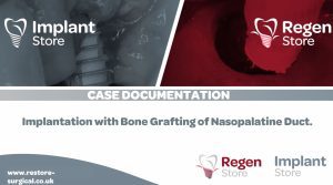
Implant and Grafting Nasopalatine duct_Dr M Leventis_Low Res


CREATED BY EXPERTS FOR EXPERTS


REFERENCES
1.Iimori Y, Kameshima Y, Yasumori A, Okada K. Effect of solid/solution ratio on apatite formation from CaSiO3 ceramics in simulated body fluid. J Mater Sci Mater Med 2004;15:1247–1253.
2.Xu S, Lin K, Wang Z, Chang J, Wang L, Lu J, Ning C. Reconstruction of calvarial defect of rabbits using porous calcium silicate bioactive ceramics. Biomaterials 2008;29:2588–2596.
3.Hing KA, Wilson LF, Buckland T. Comparative performance of three ceramic bone graft substitutes. Spine J. 2007; 7(4):475-490.
4.Nagineni, Vamsi V., et al. "Silicate-substituted calcium phosphate ceramic bone graft replacement for spinal fusion procedures." Spine 37.20 (2012): E1264-E1272.
5.Dashnyam, K.; El-Fiqi, A.; Buitrago, J.O.; Perez, R.A.; Knowles, J.C.; Kim, H.-W. A mini-review focused on the proangiogenic role of silicate ions released from silicon-containing biomaterials. J. Tissue Eng. 2017, 8, 1–13. 
6.Test Report Bonegraft Biologic, No: 2018-BME-05-1, 2018-BME-05-2, 2018-BME-05-3 and 2018-BME-05-4.
7.Licina et al, Comparison of (SiCAP) with (Infuse) in Posterolateral Instrumented Lumbar Fusion Global Spine J 2015;5:471–478.
9.Wheeler, et al. Efficacy of silicated calcium phosphate graft in posterolateral lumbar fusion in sheep. The Spine Journal 7 (2007) 308–317

Clinical Images: Dr Minas Leventis.